Advanced Imaging

An Introduction

From day-to-day workhorses to super-resolution imaging, we have all your imaging needs covered. We work with you on experimental design, guide you to the appropriate instrument, provide training and assisted use on our scopes, and help with image and data analysis. The NBCC is partnered with the Nikon Centre of Excellence at the Lunenfeld-Tanenbaum Research Institute, which provides our users with early access to advanced instrumentation and data analysis workflows.

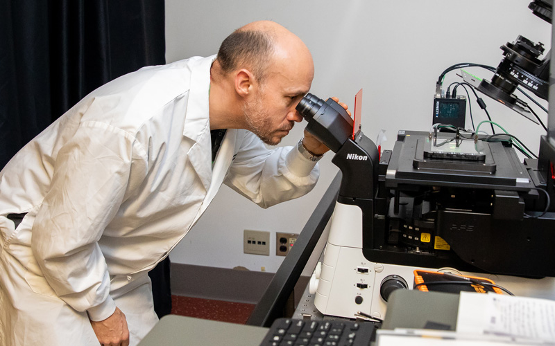
Johnny looking into a microscope

Quick Navigation

Services

1. Super-Resolution Imaging

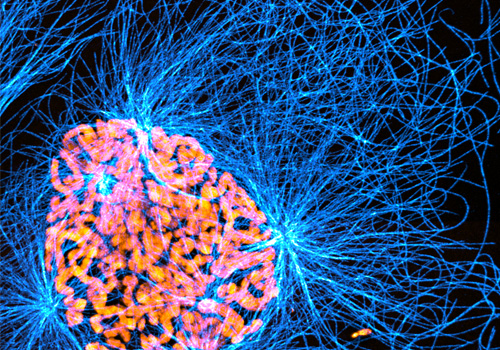
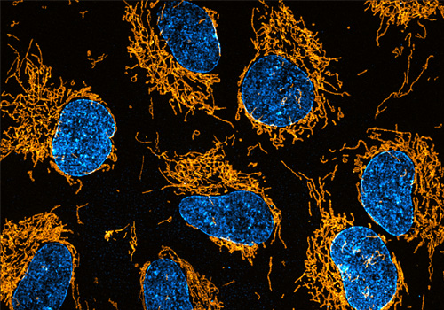
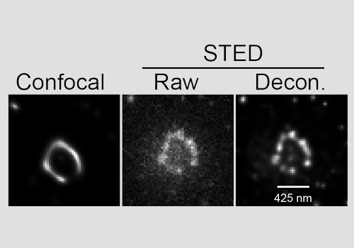
Super-Resolution imaging goes beyond the diffraction limit of light to provide more detailed analysis of sub-cellular structures. We offer three different super-resolution options. The SoRa and DeepSIM systems provide up to 100 nm resolution with standard fluorophores in fixed or live samples. The STEDYCON system can achieve 30 nm resolution for the ultimate detail.

SoRa - RPE-1

SoRa - RPE-1 cell in Prophase

DeepSim - RPE-1 cells

DeepSim - RPE-1 cells stained with Tomm20 antibody

ANKRD26 in RPE-1 cells using STEDYCON

Deconvolution of ANKRD26 in RPE-1 cells using STEDYCON


2. Dynamic Live-Cell Imaging

Our widefield DeltaVision system is ideal for live-cell imaging using fluorescently-tagged molecules or live cell compatible dyes. Temperature, humidity and CO2 control are available for long-term live cell imaging to reveal molecule dynamics.


3. Whole-Slide Scanning

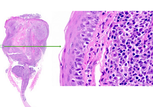
Our Zeiss Axio Z1 slide scanner will allow up to 100 slides to be imaged with either a Brightfield or Fluorescent camera, allowing the whole slide to be digitized at either 20x or 40x, giving a full picture of the entire specimen.

Slide Scanner

4. Widefield and Confocal Imaging

We also offer a wide range of widefield and confocal microscopes to meet your imaging needs. All our instruments are detailed under equipment.


5. Image Analysis

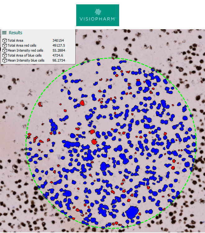
We offer the use of Visiopharm, NIS Elements, Volocity, CellProfiler, and ImageJ software. We provide training, advice and support for all your image analysis needs.

Analysis Vis

Equipment

1. Super-Resolution Instruments:

Nikon CSU-SoRA:

The Nikon CSU-SoRa is a spinning disk microscope with optical reassignment. It offers super resolution (~100 nm) of live or fixed samples with good fluorescence intensity. Its field of view is on the scale of a single mammalian cell, and it can accommodate standard secondary fluorophores (e.g., AlexaFluor), live dyes and fluorescent proteins.


Nikon CREST v3 DeepSIM

The Nikon CREST v3 DeepSIM is a spinning disk microscope with structured illumination. It offers super resolution (~ 100 nm) of live or fixed samples with a field of view that can accommodate multiple mammalian cells. Since it is less phototoxic than the SoRa, it is ideal for longer term super resolution live imaging. This scope can accommodate standard secondary fluorophores (e.g., AlexaFluor), live dyes and fluorescent proteins.


Abberior STEDYCON

The Abberior STEDYCON is a super resolution microscope with stimulated emission depletion. It offers super resolution (~30 nm) of fixed samples with a field of view up to 90 x 80 um. This system works best with Abberior STAR dyes.

Nikon CSU-SoRa Nikon CREST v3 DeepSIM Abberior STEDYCON
Objectives
  • 10x PlanApo lS 0.45 NA, air
  • 20x LWD Apo lS WI 0.95 NA, water 60x PlanApoIR W 1.27 NA, water
  • 60x PlanApoVC WI 1.20 NA, water
  • 60x ApoTIRF, 1.49 NA, oil
  • 100x ApoTIRF, 1.49 NA, oil
  • 10x PlanApo lD 0.45 NA, air
  • 20x PlanApo lD 0.8 NA, air
  • 20x LWD Apo WI lS 0.95 NA, water
  • 60x SRPlanApoIR WI 1.27 NA, water
  • 60x PlanApo lD 1.42 NA, oil
  • 100x PlanApo lD 1.45 NA, oil
  • 10x PlanApo lD 0.45 NA, air
  • 20x PlanApo lD 0.8 NA, air
  • 20x LWD Apo WI lS 0.95 NA, water
  • 60x SRPlanApoIR WI 1.27 NA, water
  • 60x PlanApo lD 1.42 NA, oil
  • 100x PlanApo lD 1.45 NA, oil
  • Excitation wavelengths
  • 405 nm (DAPI)
  • 445 nm (CFP)
  • 488 nm (GFP)
  • 514 nm (YFP)
  • 561 nm (Cy3, mScarlet)
  • 594 nm (mCherry)
  • 647 nm (Cy5)
  • 405 nm (DAPI)
  • 440 nm (CFP)
  • 488 nm (GFP)
  • 514 nm (YFP)
  • 561 nm (Cy3, mScarlet)
  • 640 (Cy5)
  • 730 (Cy7)
  • 405 nm (DAPI, non-STED)
  • 488 nm (GFP, STED only with long-stokes shift)
  • 561 nm (STAR Orange, STED)
  • 640 nm (STAR Red, STED)
  • STED Depletion N/A N/A 775 nm pulsed For use with ‘orange’ and ‘red’ or long stokes shift ‘green’ fluorophores
    Detector
  • Hamamatsu ORCA-Fusion BT sCMOS camera
  • Back illumination
  • 21.2 mm diagonal field of view
  • 6.5 mm pixel size
  • 95% peak quantum efficiency
  • 400 fps
  • Teledyne Kinetix sCMOS camera
  • Back illumination
  • 29.4 mm diagonal field of view
  • 6.5 mm pixel size
  • 96% peak quantum efficiency
  • 498 fps
  • 775 nm pulsed For use with ‘orange’ and ‘red’ or long stokes shift ‘green’ fluorophores
    Environmental Control Tokai Hit stage top incubator for control of temperature, CO2 and humidity. Tokai Hit stage top incubator for control of temperature, CO2 and humidity. Tokai Hit stage top incubator for control of temperature, CO2 and humidity.
    Associated Software NIS Elements NIS Elements Abberior acquisition software

    2. Confocal Instruments:

    We have a Nikon A1 point scanning confocal, a Leica Quorom spinning disc confocal, and a BC43 spinning disc, widefield scope in the facility

    Nikon A1 Leica Quorom BC43
    Location Hospital rm 865 Orde 6-1001-3 (Microscope Room B) Orde 6 (Santos Lab)
    Model Nikon Leica Andor
    Microscope Leica DMI 6000 inverted, motorized BC43 - Spinning Disc and Widefield
    Lenses
  • 4X
  • 10X
  • 20X
  • 40X
  • 63X
  • 5x Nplan 0.12 NA, air
  • 10x HCPLApo 0.40 NA, air
  • 20x HCPlan Apo 0.70 NA, air
  • 40x HCx PlApo 1.25 NA, oil
  • 63x HCx PlApo 1.32 NA, oil
  • 100x HCx PlApo 1.40 NA, oil
  • 2x 0.06 NA, air
  • 10x 0.45 NA, air
  • 20x 0.75 NA, air
  • 40x 0.75 NA, air
  • 63x 1.4 NA, oil
  • Vibration isolation Yes Yes Yes
    Stage Motorized xyz, Slide/Plate/dish holder, Software-integrated Motorized xyz, Slide/Plate/dish holder, Software-integrated Motorized xyz, Slide/Plate/dish holder, Software-integrated
    Z-focus Microscope Microscope or piezo Microscope and autofocus
    Illumination
  • Lasers 405,488,561,647
  • Halogen 12V 100W
  • EXFO X-Cite 120
  • Lasers 405,488, 561 and 647
  • Lasers 405,488,547,633
  • Filtering
  • b) DAPI 460/50
  • FITC 525/50
  • Cy3 ff593/40
  • CFP 470/24
  • TxRed 620/60
  • Cy5 700/75
  • b) eye piece viewing filter cubes (DAPI/GFP/RFP)
  • c) DIC
  • DAPI
  • FITC
  • Cy3
  • Cy5
  • Detector Hamamatsu C9100-13 EM CCD Camera
    Pinhole Adjustable Fixed Fixed
    Software Elements Volocity Fusion

    3. Widefield Instruments:

    We offer 5 different widefield instruments from a DMIL LED for basic histology and fluorescence to a DeltaVision Elite for long-term imaging.

    DeltaVision BX61 – fluorescent and histology Olympus IX81 Inverted (OPTIGRID) BX61 – histology only DMIL LED
    Location 1065 865 (SW) Orde 6-1001-2 (Microscope Room A) Orde 6-1001-2 (Microscope Room A) Orde 6-1014-1 (Tissue Culture Microscope)
    System Type WF/Deconvolution (200 nm resolution) histo-fluor (camera-based) WF/Structured Illumination and Deconvolution Histology, colour camera Basic Histology and WF fluorescence, colour camera
    Model Elite Olympus OptiGrid on IX81 and Huygens Deconvolution Olympus Leica inverted, basic, photography and simple measurements/image processing
    Microscope Olympus IX-71, inverted Olympus BX61 Olympus IX81 inverted Olympus BX61 DMIL LED
    Lenses
  • 4x UPlanFLN 0.13 NA, air
  • 10x UPlanApo 0.4 NA, air
  • 20x UPlanApo 0.75 NA, air
  • 20x UPlanApo 0.85 NA, oil
  • 40x UPlanSApo 0.95 NA, oil
  • 60x UPlanApoN 1.42 NA, oil
  • 100x UPlanSApo 1.4 NA, oil
  • 4x UPlanSApo 0.16 NA, air
  • 10x UPlanSApo 0.40 NA, air
  • 20x UPlanSApo 0.75NA, air
  • 40x UPlanFLN 1.30 NA, oil
  • 60x PlanApoN 1.42 NA, oil
  • 100x UPlanSApo 1.40 NA, oil
  • 10x air UPlanSApro 0.4NA, 20x air UPlanSApro 0.75 NA,
  • 40x air UPlanSApro 0.95 NA , 40x oil UPlanFL N 1.3 NA ,
  • 40x oil UPlanFL N 1.3 NA ,
  • 60x air UPlanSPApo 1.45 NA,
  • 100x air UPlanSApo 1.4 NA
  • Dry 2/0.08, 10/0.4, 20/0.75, 40/0.95
  • Oil 60/1.35, 100/1.4
  • Dry 4/0.10, PH10/0.25, PH20/0.35, PH40/0.55 corrected
  • Vibration Isolation Yes No No No No
    Stage Motorized x,y,z Manual xy, Slide spring clip Motorized xyz, Slide/Plate holder, Software-integrated Motorized xyz, Multislide holder, Software-integrated Manual xyz, slide, dish holder
    Z-Focus Software and hardware-based autofocus Integrated objective z-drive microscope and autofocus microscope and autofocus capable manual not integrated
    Illumination InSight SSI 7colour solid state illumination EXFO X-Cite 120 a) Halogen 12V 100W Hallogen 12V100W EXFO X-Cite 120 and 5W LED
    Filtering
  • 390 nm (DAPI)
  • 438 nm (CFP)
  • 475 nm (GFP)
  • 513 nm (YFP)
  • 542 nm (Cy3, mScarlet)
  • 575 nm (mCherry)
  • 632 nm (Cy5)
  • DAPI
  • FITC
  • TxRed
  • CFP
  • YFP
  • BF-brightfield
  • b) EXFO X-Cite Exacte
  • DAPI/FITC/TR triple bandpass
  • FITC
  • Detector SCMOS
  • a) Fluor camera: Hamamatsu DP71 camera
  • b) Color camera: Olympus DP71
  • a) filter cubes: 377/447, 436/480, 482/536, 500/535, 562/624, 620/700
  • b) triple bandpas (blue/green/red) for eyepiece viewing
  • c) DIC
  • DIC and BF
  • MicroPublisher 5.0 RTV Colour Camera
    Software
  • Volocity / Xp
  • DP Controller
  • Hamamatsu OrcaR2 CCD Camera
  • CCD Colour Camera Olympus DP72
  • QCapture Pro 6.0 /Win XP 32
    Offline analysis SoftWorx Volocity Volocity/ N/A Same Computer
    Other Features Fully enclosed stage for control of temperature, CO2 and humidity.
  • Volocity Visualization, Quantitation, and Restoration, Huygens deconvolution and Imaris modules/Win7 64/td>
  • multi point acquisition, multi frame scan,
  • Olympus CellSens
  • Vsiopharm Image analysis on virtual slides from any scanner
  • The highest BF resolution with 1.4NA oil condenser

  • 4. Slide Scanner:

    Zeiss AxioScan
    Location 865
    System Type Slide Scanner - Fluorescent and Brightfield
    Model Zeiss
    Microscope Zeiss AxioScan1
    Lenses
  • 5x Fluar 0.25NA,
  • 20x PlanApo 0.8NA,
  • 40xPlanApo 0.95NA
  • Vibration isolation Yes
    Filters
  • Dapi Ex 330-375 Em 430-470
  • FITC Ex 453-485 Em 504-546
  • Cy3 Ex 540-557 Em 578-640
  • Cy5 Ex 630-650 Em 670-710
  • Aqua Ex 436 Em 465-495
  • Camera
  • Fluorescent: Orca Flash 4.0 V2 Axio Scan
  • Colour: Hitachi HV-F202 f/ Axio Scan
  • Software Zeiss Zen
    Offline Analysis
  • Zeiss Zen Software (free download),
  • QuPath (free download),
  • Visiopharm (licensed software)
  • Pricing

    Pricing is for external academic users only. Internal or industry users should contact Johnny Tkach (tkach@lunenfeld.ca) for super-resolution microscopes and Louise Brown (louise@lunenfeld.ca) for all other instruments.

    Super Resolution External Pricing ($/Hr)
    Assisted
    Nikon CSU-W1 SoRa $99.00
    Nikon Crest V3 DeepSIM $99.00
    Abberior STEDYCON $99.00
    External Pricing ($/Hr)
    Widefield & Confocal Assisted 1Unassisted
    GE Elite DeltaVision Widefield $99.00 $35.00/$20.00
    Nikon A1 confocal $99.00 $35.00
    Quorom Spinning Disc Confocal $99.00 $35.00
    Andor BC43 confocal $99.00 $35.00
    Olympus IX81 inverted $99.00 $20.00
    Olympus BX61 fluorescent/brightfield $99.00 $18.00
    Olympus BX61 brightfield $99.00 $18.00
    Leica DMIL LED Upright $99.00 $3.00

    1For time-lapse imaging on DeltaVision, reduced price during off-peak hours (weekends and 6 pm to 9 am on weeknights).

    Zeiss Axio Z1 Slide Scanner External Pricing ($/slide)
    20x brightfield $8.00
    40x brightfield $10.00
    20x fluorescent $16.00
    40x fluorescent $22.00
    External Pricing ($/hr)
    Software Assisted Unassisted
    Volocity $99.00 $12.00
    Visiopharm $99.00 $12.00
    Nikon Elements $99.00 $12.00
    Training/Data Analysis External Pricing ($/hr)
    Training/Theory/Data Analysis $99.00

    Policies

    Accessing the facility

  • Users interested in accessing the Advanced Imaging facility should contact Louise Brown (louise@lunenfeld.ca) to discuss the scope of their project. For super-resolution imaging, users should contact Johnny Tkach (tkach@lunenfeld.ca) to discuss the scope of their project.
  • All users must be trained by facility staff. No shows for training will be charged for the full session booked.

  • Instrument Booking and Usage

  • Instruments can be booked for a maximum of 3 hours per day Mon-Fri 9-5. Off hours and weekends there is no limit.

  • Users should book for the time they need and edit their bookings if finished early. **Please note that users who constantly over book may be charged for their full booking time.

  • All users must clean objectives when oil is used and leave the system/workstation clean.

  • Report all concerns regarding the system to facility staff immediately.

  • Biosafety

  • Users who plan to do live cell imaging experiments have to meet with the manager to discuss potential bio safety risks and to ensure appropriate training.
  • Users must be familiar with practices outlined in Laboratory Biosafety Guidelines (3rd edition, 2004, Public Health Agency of Canada-PHAC and enforced by the human Pathogens and Toxins act).

  • Acknowledgement

    Preferred acknowledgment for the Network Biology Collaborative Centre Advanced Imaging Facility:

    The authors wish to thank [staff name] of the Network Biology Collaborative Centre Advanced Imaging Facility (RRID: SCR_025389) at the Lunenfeld-Tanenbaum Research Institute for [service]. The facility is supported by the Canada Foundation for Innovation and the Ontario Government.

    Click here for our publication policy and preferred acknowledgement for other NBCC Facilities.