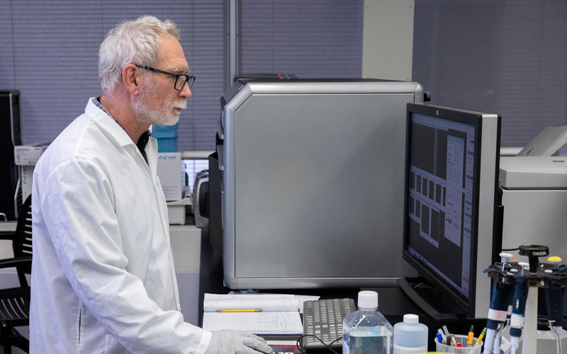
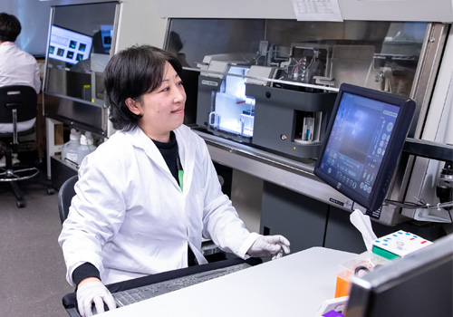
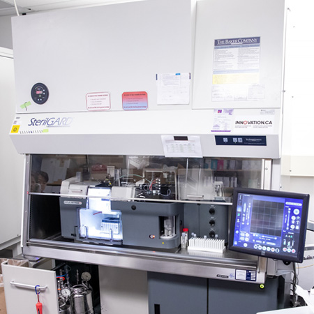
The Flow Cytometry facility offers a full complement of leading-edge instrumentation from cell-sorting and analysis to the very latest in imaging flow technology. With our specialty in image-enabled flow cytometry and cell sorting, we offer the ability to harness the combined power of imaging, traditional flow cytometry, spectral detection, and machine learning to drive discovery. We get to know our clients and their research question; then, we provide the necessary tools and expertise to assist them throughout every stage of their research from experimental design and training to data analysis and reporting.
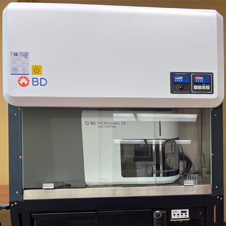
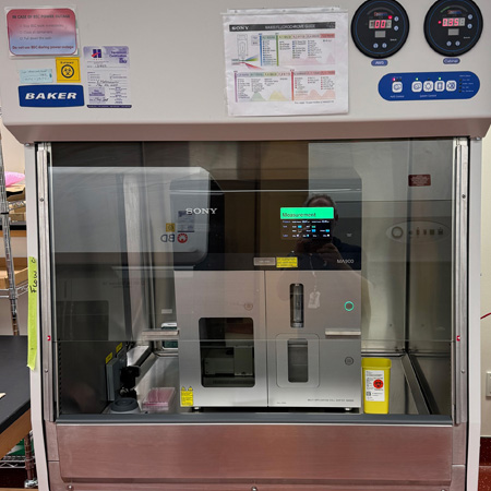
We support a variety of full-service cell sorting applications using either conventional or spectral detection for single cell or bulk sorts. Our BD FACSDiscover S8 allows for the inclusion of six additional imaging channels complexed with spectral detection to provide novel cell sorting parameters. For clients wishing to perform their own sorts, we provide comprehensive cell sort training on our Sony MA900 cell sorter(refer to the Equipment section for instrument configurations).
For information on how to prepare samples for cell sorting, refer to our sample preparation guidelines shortform, longform, BD pre sort buffer and please note that cell-sort requests must be accompanied by a biosafety form prior to booking.
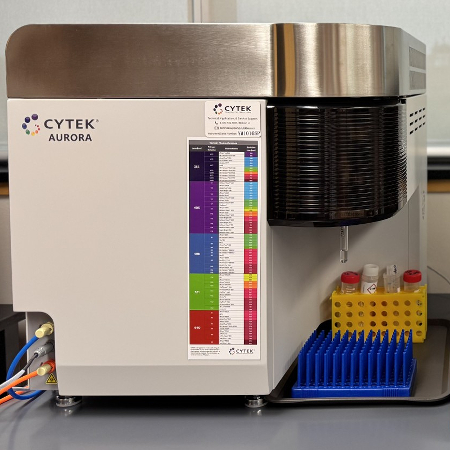
Our facilities have a full suite of conventional and image-based flow cytometry analyzers with high-throughput capability. Please refer to the Equipment section for specific instrument configurations.
For researchers who wish to perform their own flow experiments, we provide comprehensive consultation services that include in-depth theory review, experimental design recommendations, hands-on instrument training with follow-up troubleshooting, data analysis and reporting assistance.
Location | 25 Orde St, Room L421 |
Excitation | 349 nm, 405 nm, 488 nm, 561 nm, 637 nm |
Detectors | full spectrum – 78 APD fluorescence |
LL+ Scatter Detectors |
violet laser - axial light loss, SSC
blue laser (image) – FSC, SSC & Axial light loss |
Image Detectors | 488 nm excitation - fluorescence (PMT FL1 LP505:534/46 - FL2 LP570:600/60 – FL3 LP675: 788/225) |
Imaging Features | center of mass X/Y, correlation, delta center of mass, diffusivity, eccentricity, max intensity, moment long/short, radial moment, sixe, total intensity |
Nozzles | 85 mm, 100 mm, 130 mm |
Sort | 4-way, indexed, plate |
Location | 25 Orde St, Room L421 |
Excitation | 355 nm, 405 nm, 488 nm, 561 nm, 642 nm |
Detectors | 21 (355 nm/3, 405 nm/3, 488 nm/7, 561 nm/4, 642 nm/4) |
Nozzles | 70 mm, 100 mm,130 mm |
Sort | 6-way, indexed, mixed mode, plate |
Location | 25 Orde St, Room L421 |
Excitation | 405 nm, 488 nm, 561 nm, 638 nm |
Detectors | 12 (co-linear 488 nm & 561 nm/5 + co- linear 405 nm & 638 nm/7) |
Nozzles | 70 mm, 100 mm,130 mm |
Sort | 6-way, indexed, mixed mode, plate |
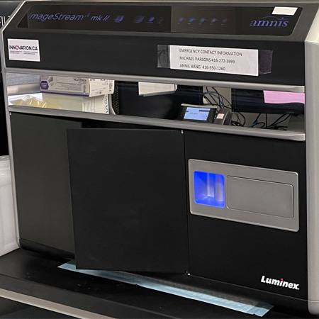
Loacation | 25 Orde St, Room L421 |
Excitation | 405 nm, 488 nm, 561 nm, 592 nm, 638 nm |
Imaging Channels | 12 channels (brightfield, darkfield + 10 fluorescent) |
Features | Upgraded camera (high gain), Upgraded 488 nm laser (400 mW), 96-well plate autosampler, DEAS v6.4 - machine learning enabled for enhanced image analysis, Amnis AI stand alone deep learning analysis software available |
Location | 600 University Ave, Room 980 |
Excitation | 355 nm, 405 nm, 488 nm, 561 nm, 640 nm |
Colours | 18 (355 nm/2 405 nm/6, 488 nm/2, 561 nm/5, 640/3) |
Features | High-throughput sampler |
Location | 600 University Ave, Room 980 |
Excitation | 405 nm, 488 nm, 561 nm, 640 nm |
Colours | 14 (405 nm/5, 488 nm/2, 561 nm/4, 640/3) |
Features | High-throughput sampler |
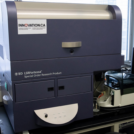
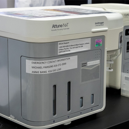
Location | 600 University Ave, Room 980 |
Excitation | 405 nm, 488 nm, 561 nm, 637 nm |
Colours | 14 (405 nm/6, 488 nm/2, 561 nm/3, 640/3) |
Features | Acoustic focusing, CytKick Max autosampler (96-, 384-well plates + 24 x 1.5 ml micro-tube rack with cooling |
Location | 25 Orde St, Room L421 |
Excitation | 405 nm, 488 nm, 561 nm, 640 nm |
Colours | 10 (co-linear 488 & 561 nm/5, 405 nm/2, 638 nm/3) |
Features | 32 tube carousel |
Instrument | Internal Pricing ($/hr) | External Pricing ($/hr) | ||
---|---|---|---|---|
Assisted | Unassisted | Assisted | Unassisted | |
BD S8 cell sorter | $100.00 | $75.00 | $115.00 | $85.00 |
MoFlo ASTRIOS cell sorter | $90.00 | n/a | $100.00 | n/a |
Sony MA900 cell sorter | $90.00 | $60.00 | $100.00 | $65.00 |
Amnis ImageStream analyzer | $90.00 | $65.00 | $95.00 | $70.00 |
Thermo Attune NXT analyzer | $85.00 | $45.00 | $90.00 | $50.00 |
Beckman Gallios analyzer | $85.00 | $45.00 | $90.00 | $50.00 |
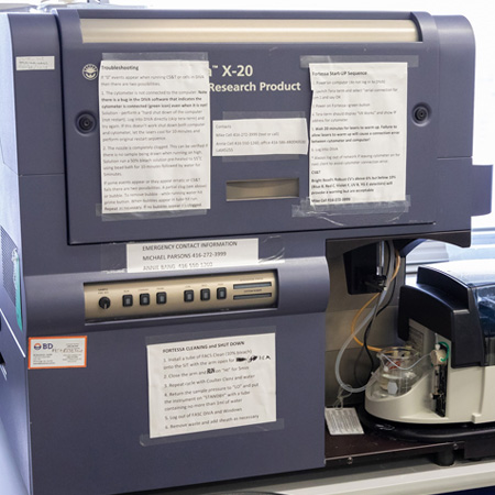
BD Fortessa X-20 analyzer | $85.00 | $45.00 | $90.00 | $50.00 |
BD Fortessa analyzer | $85.00 | $45.00 | $90.00 | $50.00 |
Training and Theory | $90.00 | n/a | $99.00 | n/a |
Data Analysis | $90.00 | $0.00 (1) | $99.00 | $0.00 |
MA900 cell sorter chip | $50.00 | $50.00 ea. | $55.00 | $55.00 ea. |
(1) There is no charge for unassisted use of computer workstations and data analysis software.
A biosafety questionnaire must be completed by all new clients and approved by facility staff before work commences and all users must review these guidelines:
Preferred acknowledgment for the Network Biology Collaborative Centre Flow Cytometry Facility:
The authors wish to thank [staff name] of the Network Biology Collaborative Centre Flow Cytometry Facility (RRID: SCR_025384) at the Lunenfeld-Tanenbaum Research Institute for [service]. The facility is supported by the Canada Foundation for Innovation and the Ontario Government.
Click here for our publication policy and preferred acknowledgement for other NBCC Facilities.