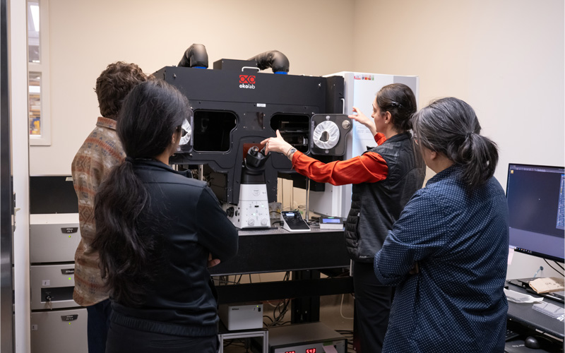
The High-Content Screening facility provides access to cutting edge high-throughput imaging equipment,
as well as powerful automated image and data analysis tools. This unit is a natural end point for
automated assays including large-scale RNAi/chemical screens. Our expertise in high-throughput imaging
and high-content analysis ensures the most efficient and robust screen design. The available microscopes
enable high-speed automated imaging of multi-well plates and regular microscopy slides at either low or
high resolution. Combined with advanced image analysis tools, this allows for the development of a wide
range of standard and easily customized applications applicable to multiple areas of biomedical
research.
Quick Navigation
Services
1. High-Content Imaging
High-Content Screening is automated image acquisition and analysis in high-throughput. It enables fast testing of many
biological conditions including arrayed genetic and chemical screens, while ensuring reproducibility and objective
quantification of images. A wide variety of different applications can be accommodated including cell counting and
morphology, colony growth tracking, drug screening, cell viability and proliferation assays, protein translocation, and
co-localization assays. Assays can be performed in multi-well format on fixed or live cells in monolayers or organoids.
Time-lapse imaging for multiple plates using plate scheduler modules is available on the BioPipeline (44 plates) and the Incucytes (6 plates).
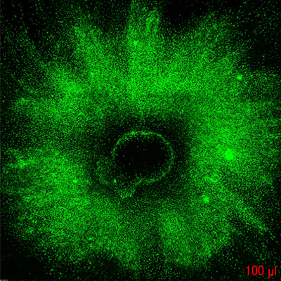
BioPipeline whole cerebral organoid embedded in a vascularized hydrogel, 4x, FITC (A. Yamand, Wrana lab, LTRI)
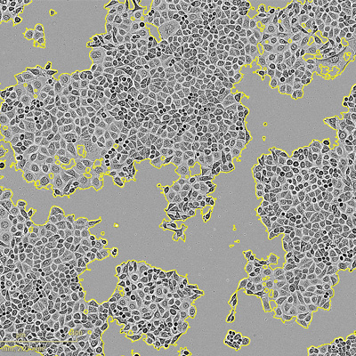
Incucyte confluency assay, phase with segmentation mask (K. Pacholczyk, Swallow lab, LTRI)
IN Cell Scratch Wound Healing Assay - 4x magnification, phase contrast (D. Ng, Swallow Lab, LTRI)
2. High-Content Data Analysis
We have several tools for automated image analysis. They include ones which can be used without extensive training to create
simple analysis algorithms (Incucyte Analysis, Celígo Analysis and Columbus), as well as software.
NIS-Elements HC (Nikon)
NIS-Elements HC is a total acquisition-to-analysis solution for high-content imaging applications. It streamlines high-speed,
automated multi-well plate acquisition, data review, analysis and management of multiple plate experiments. Multiple
scans can be analyzed simultaneously during the imaging phase or run post-acquisition on offline stations. Heat maps of well/plates,
sample images, binary masks, assay results, sample labels and other metadata are centralized for quick filtering, gating and
drill down to cellular detail. NIS-Elements offers histogram, scatterplot, bar chart, XY line, classification and gating
functions. Users can easily navigate within the Plate View and export to Excel or bitmap.
Celígo Analysis (Nexcelom Bioscience)
This built-in image analysis tool allows on-the-fly analysis of acquired images. Recommended applications include label-free cell
growth tracking, cell cycle analysis, cell viability and apoptosis, transfection optimization, multiplex fluorescent expression
assays, and many others. Once image acquisition is complete, data can be re-analyzed at an offline workstation. This instrument
is ideal for up to 4-channel imaging of multi-well plates at 4X low-resolution.
Columbus (PerkinElmer)
Columbus is a universal image data storage and analysis system that enables access to stored images via a standard internet browser.
The Columbus system allows scientists to remotely access, view, annotate, and analyze images. Columbus is based on an OMERO server and
therefore supports a variety of existing microscope file formats. Integrated image analysis tools are intuitive and efficient. A
high-performance computing server enables hyper-threading of image analysis. PhenoLOGIC™ machine learning technology provides an easy
way to classify cells or subcellular regions within a given data-set.
Acapella (PerkinElmer)
Acapella is a powerful tool for image analysis. It has a set of proprietary scripts for detection of typical cell shapes and
sub-cellular organelles. This is combined with a versatile programming environment to create customized algorithms for a
variety of biological applications.
Matlab (Mathworks)
Matlab is the most powerful application for advanced analysis of images. Flexible image and data handling allows highly specialized
routines for image analysis, including complex image filtering and segmentation. Costing for this analysis will be determined based on
the complexity of the analysis.
Equipment
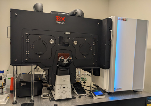
1. Nikon BioPipeline
The Nikon BioPipeline
The Nikon BioPipeline is a high-end CSU-W1, dual camera, dual spinning disk confocal
high-content platform with 7 imaging channels. It is based on Nikon's Eclipse fully
motorized, inverted Ti1 microscope and the A1R-HD25 system. An integrated 44-multi-
well plate incubator provides multi-user assay scheduling. It is ideally suited for high-
resolution fixed or live screening assays such as detection of DNA damage foci,
translocation assays, measuring of cilia lengths, co-localization studies, drug screenings,
or any other assay that needs to combine high-resolution with automated imaging.
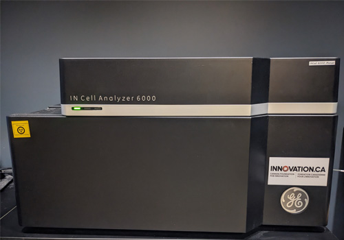
2. IN Cell Analyzer 6000
The IN Cell Analyzer 6000 is a laser-based, line-scanning confocal imaging platform with both widefield and confocal capabilities. This instrument is
designed for high-throughput fixed or live imaging and assay development on a variety of vessel types. It can be used for timelapse and endpoint assays
as well as 3-D imaging, co-localization studies and low signal assays.
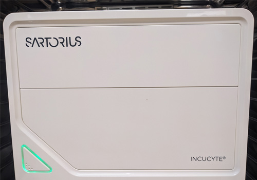
3. Sartorius Incucytes
The Incucyte platforms are automated, high-content, widefield, multi-well plate acquisition, analysis and visualization platforms designed for long live
imaging within an incubator. They offer dynamic insights into the health, viability, morphology, movement and function of cell models, with assays
including confluence monitoring, spheroid and organoid detection, and scratch-wound. Our facility has an Incucyte S3 and the latest Incucyte SX5.
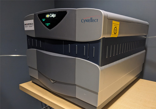
4. Celígo
Celígo is a low-resolution high-throughput microscope, equipped with LED-based light sources (brightfield and 3 fluorescent channels). Uniform well
illumination and large-chip CCD camera allow users to image entire wells at a high-speed. Built-in image analysis tools allow performing on-the-fly
analysis in a variety of biological applications (cell/colony counting, migrations assays, expression analysis and many others).
Specification
Nikon BioPipeline Live
In Cell Analyzer 6000
Incucyte
Celigo
Type
Spinning-disk confocal
Line-scanner confocal, widefield
Widefield
Widefield
Objectives
• 4X Nikon Plan Apo 0.2 NA 20 mm WD, Air
• 4X Nikon Plan Fluor Phase Contrast 0.13 NA 17.1 mm WD, Air
• 10X Nikon Plan 10X 0.45 NA 4 mm WD, Air
• 10X Nikon Plan Fluor Phase Contrast 0.3 NA 0.16 mm WD, Air
• 20X Nikon Plan Apo 0.75 NA 1 mm WD, Air
• 20X Nikon Apo Water 0.95 NA 0.95 mm WD, Water
• 20X Nikon S Plan Fluor ELWD 0.45 NA
• 20X Nikon Super Plan Fluor Phase Contrast ELWD 0.45 NA 6.9-8.2 mm WD, Air
• 40X Nikon Plan Apo 0.95 NA 0.17-0.25 mm, Air
• 40X Nikon Apo 1.15 NA 0.59-0.61 mm WD, Water
• 40X Nikon S Plan Fluor ELWD 0.4 NA 3.6 mm WD, Air
• 40X Nikon Super Plan Fluor Phase Contrast ELWD 0.6 NA 2.8-3.6 mm WD, Air
• 60X Nikon Plan Apo 1.2 NA 0.28-0.31 mm WD, Water
• 60X Nikon Plan Fluor Air ELWD 0.7 NA 1.8-2.6 mm WD, Air
• 60X Nikon Super Plan Fluor Phase Contrast ELWD 0.7 NA 1.8-2.6 mm WD, Air
• 4X Nikon Plan Apo 0.2 NA 20 mm WD, Air
• 10X Nikon Plan Apo 0.45 NA 4 mm WD, Air
• 20X Nikon Plan Fluor ELWD 0.45 NA 7.5 mm WD, Air
• 60X Nikon Plan Fluor ELWD 0.7 NA 1.8 mm WD, Air
• Blue 405 nm
• Green 488 nm
• Red 561 nm
• Far-red 640 nm
• 1st module:
• Green 440-480 nm (Incucyte S3, SX5)
• Red 565-605 nm (Incucyte S3, SX5)
• Swappable module Incucyte
• Green 453–485 number
• Orange 648-674 nm
•NIR 576–639 nm
• Blue 377+/-50 nm
• Green 483+/-32 nm
• Red 531+/-40 nm
• Far-red 628+/- 40 nm
Non-fluorescent Channels
Brightfield, Phase
Brightfield, DIC, Phase
Phase
Brightfield
Imaging Modality
2D, 3D
2D, 3D, Maximum intensity projection
2D
2D
Environmental Control
Yes
Yes
Yes
No
Associated Software
NIS Elements
Columbus
Built-in Incucyte analysis
Built-in Celigo analysis
Pricing
Pricing is for external academic users only. Internal or industry users should contact Monica Hasegan (hasegan@lunenfeld.ca) for pricing.
HCS Instruments
Assisted
First 5 hours1
Next 5 hours
After 10 hours
Nikon BioPipeline
$108.00
$44.00
$15.00
$5.00
GE IN Cell 6000
$108.00
$37.00
$12.00
$5.00
Celígo
$108.00
$25.00
$10.00
$5.00
1Discounts are based on continuous imaging. Time-on-instrument is charged until samples are removed and software is closed. All rates are per hour.
Incucytes
External Pricing ($/plate/day)
Essen Incucyte SX5
$50.00
Essen Incucyte SX3
$50.00
External Pricing ($/hr)
Software
Assisted
Unassisted
Image Analysis Workstation
$108.00
$12.00
Assisted Use
External Pricing ($/hr)
Training/Theory/Data Analysis/Assay Development
$108.00
HCS Instruments
Assisted
Unassisted (1st hour)
Unassisted (subsequent hours)
Offpeak1 (1st hour)
Off-peak1 (subsequent hours)
Nikon BioPipeline
$99.00
$75.00
$53.00
$25.00
$16.00
GE IN Cell 6000
$99.00
$75.00
$53.00
$25.00
$16.00
Celígo
$99.00
$30.00
$21.00
$10.00
$6.00
1Peak hours are Monday to Friday, 9:00 AM to 6:00 PM
Incucytes
External Pricing ($/plate/day)
Essen Incucyte SX5
$110.00
Essen Incucyte SX3
$110.00
External Pricing ($/hr)
Software
Assisted
Unassisted
Image Analysis Workstation
$99.00
$12.00
Assisted Use
External Pricing ($/hr)
Training/Theory/Data Analysis/Assay Development
$99.00
Policies
Accessing the Facility
For accessing the facility, training on an instrument, or submitting samples for imaging,
please go to our facility management platform, Infinity X.
For New Users of Infinity X
For any questions about lab or user accounts, please contact Monica Hasegan (hasegan@lunenfeld.ca).
If you are an internal Sinai Health or Lunenfeld-Tanenbaum researcher, please click
on the "Sinai Health" or "LTRI" logo to sign into your account.
If your lab is not registered, please contact Monica.
If you are an external user and this is the first time your lab is using Infinity X, please contact Monica for instructions.
Once your lab is registered, please "Register" for a new account.
After you have an account, you will need to request access to the Advanced Imaging and High-Content Screening Facility and
then submit a "General meeting or training" form or the appropriate sample submission form.
All new users must review our getting started guide.
Biosafety
Users who plan to do live cell imaging experiments have to meet with the manager to discuss potential bio-safety risks and to ensure appropriate training. Users must be familiar with practices outlined in Laboratory Biosafety Guidelines (3rd edition, 2004, Public Health Agency of Canada-PHAC and enforced by the human Pathogens and Toxins act).
Data Management
Data storage is the sole responsibility of the client and clients are urged to transfer data to their own storage devices as soon as possible. Data will be removed regularly from the instrument workstation since it adversely affects instrument performance.
Data storage on the dedicated analysis workstations will be removed when necessary (and without notice), so please use access to the LTRI network drives available on the analysis workstations to save your data or store directly to external storage devices.
Acknowledgement
Preferred acknowledgment for the Network Biology Collaborative Centre High-Content Screening Facility:
The authors wish to thank [staff name] of the Network Biology Collaborative Centre High-Content Screening Facility (RRID: SCR_025391) at the Lunenfeld-Tanenbaum Research Institute for [service]. The facility is supported by the Canada Foundation for Innovation and the Ontario Government.
Click here for our publication policy and preferred acknowledgement for other NBCC Facilities.